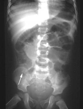
Abdominal x ray reveals a calcified round body in right lower quadrant

Plain X-ray of the abdomen showing classical curvilinear calcification

Diagnostic Approach to Benign and Malignant Calcifications in the Abdomen and Pelvis
Small-Bowel Obstruction Imaging and Diagnosis: Practice Essentials, Radiography, Computed Tomography

Abdominal x ray reveals a calcified round body in right lower quadrant

Minilaparotomy approach for biliary ileus.

PDF) Minilaparotomy approach for biliary ileus: Case report.

Presentation1, interpretation of x ray of the abdomen.
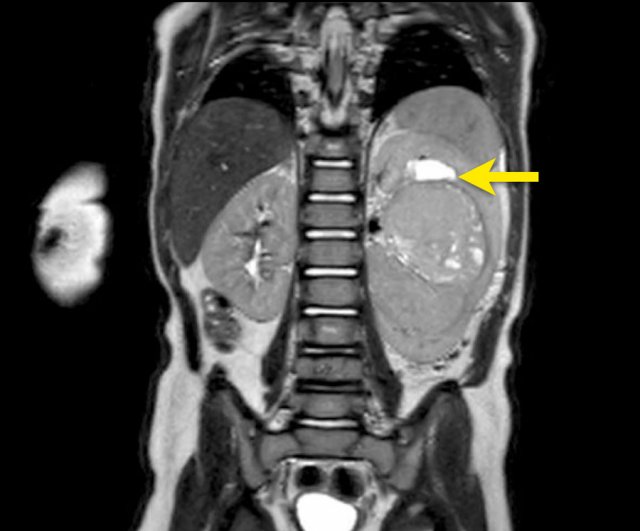
The Radiology Assistant : Solid Abdominal Masses in Children

Perforation in gall bladder at Hartman' pouch.
You may also like


Related products